Ventral and incisional hernia repair
Understand how abdominal wall weaknesses develop after previous surgery or muscular strain, and how advanced retrorectus mesh reconstruction restores lasting core stability.
What is a ventral or incisional hernia?
A ventral hernia occurs when abdominal tissue or intestine pushes through a weakness in the anterior abdominal wall muscles. When the defect develops at the site of a previous surgical incision, it is classified as an incisional hernia.
Without timely surgical repair, the defect in the muscle layer gradually enlarges due to daily breathing and day to day activities. Modern repair focuses on anatomical reconstruction: closing the midline defect and placing a prosthetic mesh in the retromuscular space where natural abdominal pressure keeps it securely anchored.
Key Diagnostic Signs & Symptoms
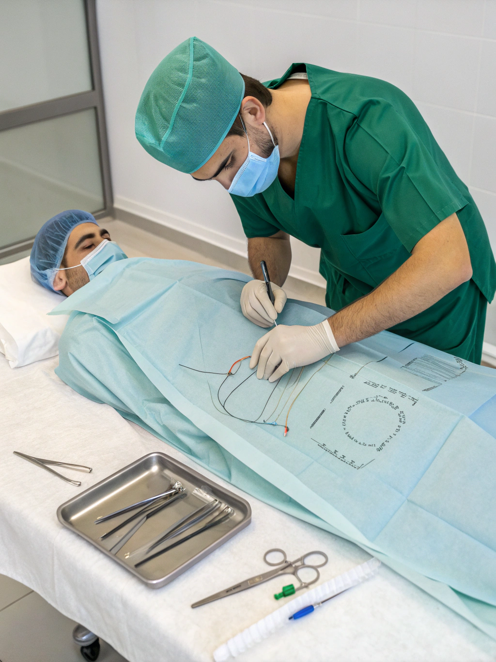
Retrorectus (Rives-Stoppa) placement isolates the surgical mesh between rectus abdominis muscle and the posterior rectus sheath, preventing direct adhesion to abdominal organs.
Underlying causes and risk factors
Ventral hernias develop when localized mechanical tension exceeds the tensile strength of the abdominal fascia.
Prior surgical incisions
Incision sites from earlier abdominal operations can weaken over time if fascia healing is compromised.
Increased intra-abdominal pressure
Chronic cough, heavy lifting, or repeated strain places constant outward stress on midline muscle layers.
Tissue healing factors
Diabetes, smoking, or wound infections during previous surgery can reduce collagen strength along the scar line.
Natural midline thinning
Gradual separation of the rectus muscles (diastasis) thins the linea alba, creating vulnerable points.
Modern tension-free repair approaches
Dr. Nitin Rimmalapudi specializes in advanced laparoscopic and robotic reconstruction techniques that reconstruct the midline rather than simply bridging the gap.
eTEP Rives-Stoppa Repair
Accesses the retrorectus space without entering the peritoneal cavity. Places a durable sublay mesh behind the rectus muscle, preventing direct bowel contact and dramatically reducing recurrence rates.
Robotic Transversus Abdominis Release (r-TAR)
For complex, wide, or recurrent incisional defects. High-definition 3D articulation allows the surgeon to release lateral abdominal layers, re-approximating the midline under minimal tension.
Have your scan reviewed by Dr. Nitin Rimmalapudi
Consult with our surgical team at Apex Hospitals or Delta Hospitals in Rajahmundry for tailored evaluation and pre-surgical planning.