How robotic hernia repair works
Robotic-assisted hernia surgery gives your surgeon enhanced vision and dexterity. Rather than working with rigid instruments, the surgeon controls precision micro-tools directly from an ergonomic master console inside the operating suite.
High-definition 3D visualization
A magnified binocular view gives the surgeon true depth perception, showing delicate tissue layers, nerves, and vessels clearly.
Articulating EndoWrist instruments
Miniature instruments rotate with greater range of motion than the human wrist, with tremor filtration for steady, measured suturing.
Sub-millimeter preperitoneal dissection
Enables precise placement of prosthetic mesh behind the muscle wall without disturbing the bowel or peritoneal cavity.
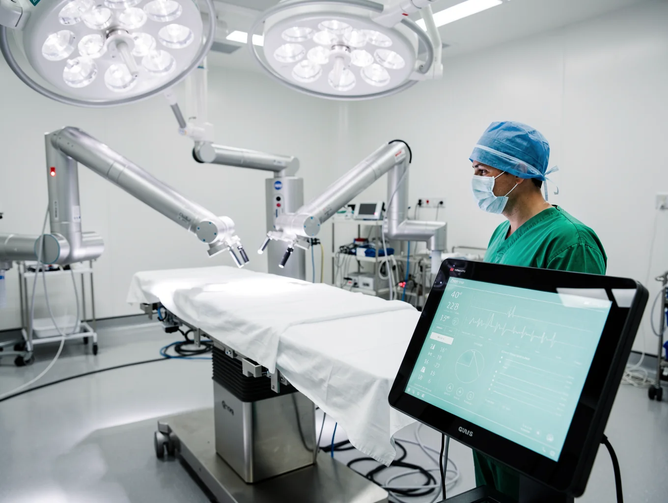
Proctored robotic cases are performed under strict safety standards with real-time console feedback and zero direct hand-tremor transfer.
Certified robotic proctor status and clinical standards
Dr. Nitin Rimmalapudi serves as a certified robotic hernia proctor and trainer, helping surgical teams across India adopt safe, standardized robotic and minimally invasive abdominal wall reconstruction protocols.
Structured surgical mentorship
Proctoring pathways combine preoperative risk evaluation, dual-console operative mentorship, and post-procedure registry follow-up to ensure clinical safety and surgical repeatability.
Pre-Operative Case Selection
Standardized CT scan imaging protocols, fascial defect measurements, and patient risk stratification before robotic console docking.
Dual-Console Mentorship
Hands-on guided dissection, retromuscular space creation, and precision articulating suture training for visiting surgeons.
Standardized Clinical Audits
Transparent registry documentation tracking operative times, pain scores, seroma incidence, and post-discharge recovery milestones.
For surgeons and referring clinicians
Inquire about robotic surgical proctoring schedules, case observation, or refer a complex ventral hernia case.
How we choose your surgical approach
Every abdominal wall is distinct. We evaluate your imaging, prior scars, and physiological fitness to match you with the safest, most durable repair.
Core diagnostic parameters evaluated during consultation
Exact measurement of the muscle separation via high-resolution CT scan.
Mapping adhesions from prior abdominal procedures and previous mesh implants.
Assessing tolerance for laparoscopic gas insufflation and anesthesia duration.
Evaluating whether component separation (TAR) is needed to close the midline.
Unsure which repair method fits your condition?
Dr. Nitin Rimmalapudi reviews your CT cross-sectional scans personally at our Rajahmundry Centre of Excellence to determine whether standard laparoscopic, eTEP, or robotic surgery delivers the lowest recurrence risk.
Technique selection is strictly individualized based on pre-operative radiological imaging and clinical assessment.